ZEISS Ocular Imaging

CLARUS
Ultra-widefield
Imaging Exhibition
A story behind every capture
June 2023
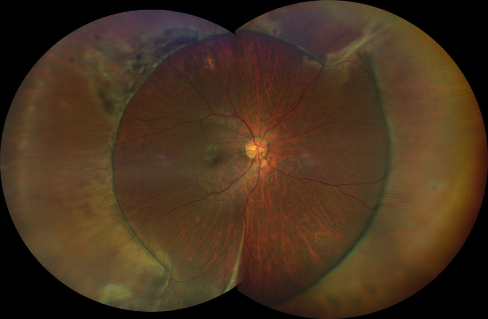
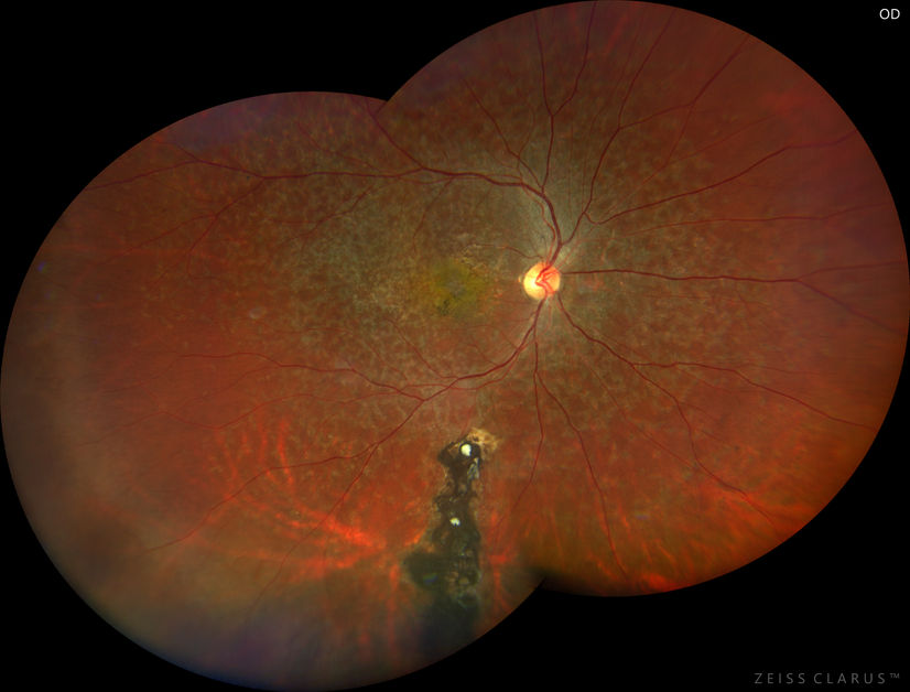
Welcome to "Clarus Ultra-Widefield Imaging Exhibition: A story behind every capture" a unique Exhibition that harmoniously weaves together the realms of medical science and artistic expression through the captivating medium of Broad Line Fundus Imaging (BLFI) in Ultra Widefield fundus photography. Join us on a remarkable journey where each fundus image serves as a canvas, combining the precision of medical understanding with the evocative power of artistry.
As we celebrate the beauty and intricacy of the human eye let these images also serve as a poignant reminder of the importance of ocular health. Through these images, may you be inspired to appreciate the marvels of our visual world and take proactive steps to preserve the precious gift of sight.
We like to express our greatest appreciation to all the submitters in this imaging competition. It is a collective effort of unveiling stories across Asia where each photograph is a testament to the resilience, fragility, and interconnectedness of human life.
Featured Images
Click to view gallery
Photos with the Winners and Judges

The CLARUS 700 ultra-widefield camera has become an essential tool in the daily practice of our multispecialty group. From eye screening, pre-refractive surgery assessement, glaucoma, and retinal disease monitoring, CLARUS fundus images are the fulcrum around which we co-manage, educate, and monitor the progress of our patients.
Asia Pacific Eye Centre

Dr. Wong Chee Wai
The ZEISS CLARUS provides pristine high fidelity true color ultra-wide field imaging, which makes interpreting diabetic retinopathy and other pathology much easier for all levels of expertise from retina specialist to ophthalmic grading technicians.
SNEC Ocular Reading Centre (SORC)

Associate Professor Gavin Tan
CLARUS 700 revolutionizes eye care as it optimizes workflow efficiency, enables exceptional care for patients without the need for indirect Ophthalmoscopy. It streamlines documentation, detects and monitors small retinal changes with precision. Doctor-patient experience is enhanced as patients can visually observe and understand their eye health better.
Larazabal Eye Clicnic

Dr. Potenciano "Yong" Larazabal III


![[Best Images Award] A Bloody Retina](https://static.wixstatic.com/media/19f4e1_b9462a416a6a4472930700284784d25e~mv2.jpg/v1/fit/w_704,h_629,q_80,enc_avif,quality_auto/19f4e1_b9462a416a6a4472930700284784d25e~mv2.jpg)
![[Best Images Award] Traffic in the Retina World](https://static.wixstatic.com/media/19f4e1_162654a05de540b981cf660bd1557ade~mv2.jpg/v1/fit/w_628,h_629,q_80,enc_avif,quality_auto/19f4e1_162654a05de540b981cf660bd1557ade~mv2.jpg)
![[Most Popular Images Award] Shimmer in the Celestial](https://static.wixstatic.com/media/19f4e1_aea46c3436404388a0749925d3159b7c~mv2.jpg/v1/fit/w_622,h_629,q_80,enc_avif,quality_auto/19f4e1_aea46c3436404388a0749925d3159b7c~mv2.jpg)
![[Most Popular Images Award] Ignite the Glowing Filament!](https://static.wixstatic.com/media/19f4e1_262a405d0ff741bd93281b34a0f09f51~mv2.jpg/v1/fit/w_1026,h_629,q_80,enc_avif,quality_auto/19f4e1_262a405d0ff741bd93281b34a0f09f51~mv2.jpg)
![[Most Popular Images Award] Unveiling the Torn Canvas](https://static.wixstatic.com/media/19f4e1_f98c7092263c4b1b9388c5e99fb621ba~mv2.jpg/v1/fit/w_629,h_629,q_80,enc_avif,quality_auto/19f4e1_f98c7092263c4b1b9388c5e99fb621ba~mv2.jpg)


